How good is prone-back position ECG to detect arrhythmias and ischemic changes in these patients? According to a recent study published in JACC Clinical Electrophysiology, prone back (PB) position ECG can serve as a useful screening tool with good diagnostic utility in COVID-19 patients who require prone ventilation.
In COVID-19 patients with hypoxic respiratory failure, early intubation and ventilation including prone-position ventilation have been recommended in society guidelines. As taking a routine supine position ECG can be cumbersome in these patients, the value of a prone position ECG needs to be explored.
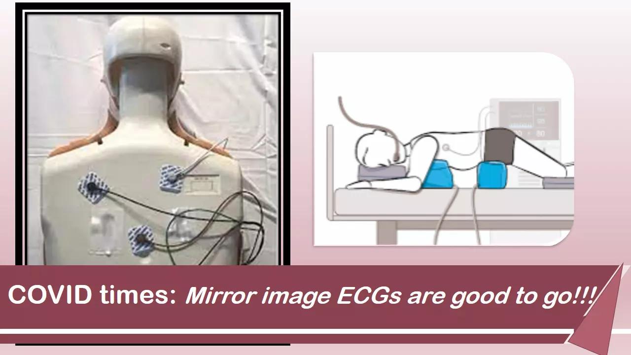
Here, the precordial leads are positioned posteriorly in a mirror image to usual anterior chest wall positions in 4th and 5th intercostal spaces (Figure 1). This avoids repositioning patients for a 12-lead ECG, which may result in oxygen desaturation and is labor-intensive.
Chieng et al conducted a study to describe expected changes in a mirror-image prone electrocardiogram (ECG) compared with normal supine, including a range of cardiac conditions. This prospective study was designed to:
1) Describe expected differences in the 12-lead ECG in the prone position compared with the supine and;
2) Determine the usefulness of the prone ECG in detecting myocardial injury and abnormalities in rhythm and conduction.
Hundred patients each underwent 3 ECGs: standard supine front (SF); prone position with precordial leads attached to front (PF); and prone with precordial leads attached to back in a mirror image to front (PB).
The study gave the following insights:
a) Prone positioning was associated with a numerically small, but statistically significant, QTc prolongation.
b) PB was associated with new qR morphology in leads V 1 to V 3 in 90% and a significant reduction in R-, S-, and T-wave amplitude in leads V 1 to V 3 .
c) In patients with anterior myocardial ischemia/infarct with ST changes in leads V 1 to V 3 on SF ECG, these changes were no longer visible in V 1 to V 3 on PB. By contrast, ST-segment elevation/depression in the limb leads remained mostly unchanged in the PB position.
d) PB positioning was associated with a change in the polarity of the T wave in leads V 1 to V 3 in 84%. T wave inversion in lateral precordial/limb leads remained unaffected by PB positioning.
e) LBBB demonstrated a polarity shift, with a RBBB appearance in a PB position.
f) RBBB became narrower with a qR appearance in V 1 to V 3 .
g) Arrhythmia detection was not affected by prone positioning.
ECG lead placement in a posterior body position is not a unique concept. Classically posterior leads V 7 to V 9 , have been used as an extension of precordial leads V 1 to V 6 in the detection of posterior myocardial infarction .
The PB ECG provides reliable rhythm monitoring and detection of myocardial injury involving the inferior and lateral myocardium, making it a useful alternative to the traditional supine ECG to avoid repositioning a patient who requires prone ventilation. However, the PB ECG is not reliable for the detection of anterior ischemia.
Machine learning–based prediction models could be developed to predict ECG appearances in a traditional supine front position based on the appearance of a prone back ECG.
Source: JACC CE: DOI: 10.1016/j.jacep.2021.04.011
Disclaimer: This website is designed for healthcare professionals and serves solely for informational purposes.
The content provided should not be interpreted as medical advice, diagnosis, treatment recommendations, prescriptions, or endorsements of specific medical practices. It is not a replacement for professional medical consultation or the expertise of a licensed healthcare provider.
Given the ever-evolving nature of medical science, we strive to keep our information accurate and up to date. However, we do not guarantee the completeness or accuracy of the content.
If you come across any inconsistencies, please reach out to us at
admin@doctornewsdaily.com.
We do not support or endorse medical opinions, treatments, or recommendations that contradict the advice of qualified healthcare professionals.
By using this website, you agree to our
Terms of Use,
Privacy Policy, and
Advertisement Policy.
For further details, please review our
Full Disclaimer.
0 Comments
Post a comment
No comments yet. Be the first to comment!